Members


About
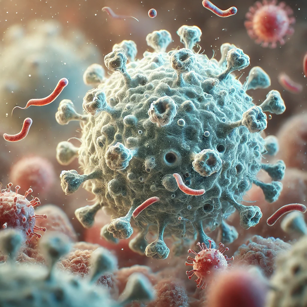
Among the myriad of immune cells, there is one subset with a unique morphology. These are dendritic cells, so named because, under the microscope, they have a star-shaped silhouette with elongated arms. These "branches" or "dendrites" offer a potential contact surface and reach far beyond those of ordinary spherical cells.
The role of dendritic cells is central to the functioning of the immune system. Capable of communicating with virtually all other cells, they coordinate the immune response (to infections, injuries, cancer, etc.) and perform a number of different functions themselves (e.g., antiviral, antitumor, or aimed at preventing autoimmunity or allergies by inducing immune tolerance to self-molecules and foods). In reality, the term "dendritic cells" covers several cell types, each specialized in a specific set of complementary or sometimes redundant functions. The signals they can detect, the cells with which they interact, or the organs in which they reside partly determine this combination of functional specialization and plasticity of each dendritic cell type.
Marc Dalod's team has embarked on a long journey into this complex world where there is still much to discover.
ALIGNING DENDRITIC CELL SUBPOPULATIONS BETWEEN MOUSE AND HUMANS TO BETTER UNDERSTAND THEIR IDENTITY AND DEDUCE THEIR FUNCTIONS
“Dendritic cells orchestrate both the immediate response, that of the “spontaneous” innate immune system, but also prepare its subsequent adaptive responses, allowing for a protective “memory” in case the system encounters the same pathogen a second time,” explains Marc Dalod. “Immunologists are currently seeking to better understand the world of dendritic cells, because deficiencies in this complex system sometimes contribute to the onset of diseases. This is the case, for example, with HIV infection, which is significantly less well controlled by our immune system than other viral infections. It is precisely this question that sparked my interest in this topic: what is not working in the immune response of dendritic cells to HIV?”
Studying cell function in the human body is never easy from a technical perspective, which is why the mouse is often the preferred model. However, for the extrapolation to be relevant, we must first ensure that the cell types being studied and manipulated—in this case, dendritic cell types—and the activation states they adopt based on the signals they receive, are functionally equivalent between the two species.
“Previously, dendritic cell heterogeneity was mainly described in mice. Nine subsets were known: three residing in organs specialized in immune functions (e.g., lymph nodes), five nested in different layers of the skin, the epidermis and dermis, and another population that only developed under inflammatory conditions. These dendritic cell subsets were identified as different from each other because they did not all produce the same antiviral factors (e.g., type I interferons, which are produced rapidly and in large quantities by plasmacytoid dendritic cells, pDCs) or because they expressed different markers on their surface,” explains Marc Dalod. However, at the time, it was difficult to distinguish between different types of dendritic cells and different activation states of the same type of dendritic cell depending on its tissue of residence, the time it was observed during an immune response and the pathophysiological context of its study.
“In humans, we didn't know as much, and the equivalence with the mouse dendritic cell system was a matter of debate. We then attempted to unify the model by studying dendritic cells in more detail, mapping the global gene expression profiles of the different mouse and human subpopulations, and comparing them unbiasedly for the expression of the thousands of genes that were found to be differentially expressed in the two datasets (in mice and humans). This allowed us to establish for the first time convincing homologies between dendritic cell subpopulations, not only between tissues within the same species, but also between mouse and human species.”
Dendritic cell populations characterized by the same genetic signature at the heart of their fundamental identity, conserved across tissues, species, and activation conditions, could then be described with unprecedented precision and were found to be equivalent between mouse spleen and skin, as well as between mice and humans, including their functional specialization, for example, the activation of white blood cells that destroy virus-infected cells or tumor cells, an activity in which conventional type 1 dendritic cells (cDC1s) excel. This led us to propose a simplified classification of dendritic cells into five cell types potentially valid for all tissues, all vertebrate species, and all pathophysiological conditions.
OPTIMIZE THE USE OF ANIMAL MODELS TO INCREASE THE LIKELIHOOD THAT THEIR LESSONS CAN BE USED IN THE LONG TERM TO IMPROVE HUMAN HEALTH
"We can now use mouse disease models to predict the most effective type and activation state of dendritic cells for protection, and then extrapolate these results to humans with the aim of therapeutically targeting their equivalents. Conversely, we can hope to improve a deficient immune response, as in the case of HIV, since we can precisely identify the weak link," enthuses Marc Dalod.
Therefore, my team seeks to understand which combinations of dendritic cell types and activation states promote protective antiviral or antitumor immunity, as opposed to immune tolerance, and how (reviews: Alexandre et al. Front Microbiol. 2014; Dalod et al. EMBO J. 2014; Vu Manh et al. Front Immunol. 2015a; Cancel et al. Front Immunol. 2019; Cheema et al. Methods Mol Biol. 2023; Ngo et al. Cell Mol Immunol. 2024). We also analyze how the functions of interferons (IFNs) and pDCs can be beneficial or detrimental, depending on the pathophysiological context (reviews: Tomasello et al. Front Immunol. 2014; Ngo et al. Cell Mol Immunol. 2024).
We combine mutant mouse lines, preclinical models of viral infections, cancer, or inflammation, as well as global or single-cell transcriptomics, multiparametric flow cytometry, and quantitative confocal microscopy, to identify the genetic modules that govern the ontogeny, activation trajectory, and functional polarization of cDC1s and pDCs, their interaction with natural killer (NK) cells and T lymphocytes, and the orchestration of these responses in time and space (Robbins et al. PLoS Pathog. 2007; Zucchini et al. Int. Immunol. 2007; Zucchini et al. J Immunol. 2008; Crozat et al. J Exp Med. 2010; Baranek et al. Cell Host Microbes. 2012; Cocita et al. PLoS Pathogens. 201 ; Vu Manh et al. Front Immunol. 2015a;2015b; Alexandre et al. J Exp Med. 2016; Tomasello et al. EMBO J, 2018; Abbas et al. Nat Immunol. 2020; Mattiuz et al. Clin Transl Immunol. 2021; Ghislat et al. Sci Immunol. 2021; Ghilas et al. iScience. 2021; Valente et al. Nat Immunol. 2023; Ngo et al. BioRxiv. 2024).
We particularly focus on determining the functions of molecules, genetic modules and regulatory signaling pathways whose expression/activity selectivity between dendritic cell types and activation states appears to be conserved during evolution, based on comparative transcriptomics between mouse and human.
DEVELOPMENT OF IN VITRO MODELS OF HUMAN DENDRITIC CELL TYPES TO ACCELERATE KNOWLEDGE TRANSFER FOR THE BENEFIT OF PATIENTS
We are also developing pipelines to differentiate human hematopoietic stem cells into authentic pDCs and cDC1s and manipulate them genetically or pharmacologically, to accelerate the translation of discoveries made in mice to benefit patients (Balan et al. J Immunol. 2014; Balan et al. Cell Rep. 2018; Luo et al. BioRxiv. 2023).
News

L’équipe de Marc Dalod du CIML démontre que certaines cellules immunitaires spécialisées peuvent être néfastes plutôt que protectrices lors d’infections […]

Dans le cadre d’Octobre Rose, les recherches menées par Marc Dalod, directeur de recherche au CNRS et responsable d’équipe au […]
Projects
Project: DECITIP Plasmacytoid dendritic cells (pDCs) excel in the production of type I and III interferons (IFN), […]


Project: pDCComp Individuals infected with viruses or suffering from inflammatory diseases develop a set of behavioral disorders […]

Project: PLBIO 2024 Coordinator: Thierry Walzer, CIRI, Lyon. Other participant: Nathalie Bendriss-Vermare, CRCL, Lyon. Abstract: Natural Killer cells […]

Project: ACLAMADABREC Partly through their effectiveness in activating natural killer (NK) cells and CD8 T lymphocytes […]

Project: MITI Other participants: Magali Richard, LIG, Grenoble; Lionel Spinelli, CB2M hub of CIML. Summary: This project aims to […]

Project: UNFIRE Other participants: Ciphe (Ana Zarubica), Hospices Civils de Lyon and CIRI (Sophie Trouillet-Assant, Thierry Walzer), CoVir team […]


Project: PLBIO-25 Coordinator: Marianne BURBAGE, Institut Curie, Paris. Other participant: Andrew GRIFFITHS, ESPCI, Paris. The effectiveness of CD8 T lymphocytes […]

Project: DCFUNDEVSCREEN Abstract: Studies in mice have shown the key antiviral/tumor role of plasmacytoid dendritic cells (pDCs) […]

Project: RIPIREVI Other participants: CIPHE (Ana Zarubica and colleagues). Abstract: Type I interferons (IFN-Is) are essential for […]


Project: NRegSkin Project leader: Mei LI (IGBMC, Strasbourg). Other participants: Katia Boniface, Julien Seneschal (University of Bordeaux). Summary: The skin […]

Project: REVOLUTION Coordinator: Edouard Sage, Foch Hospital, Suresnes. Other partners: Isabelle Schwartz-Cornil, INRAE, Jouy-en-Josas; Jérémie Pourchez, Ecole des Mines […]

Project: ICARO Current acellular vaccines are mostly effective against extracellular pathogens, but fail to induce a response […]


Project: PLBIO 2021 Coordinator: Christophe CAUX, CRCL, Lyon. Other participants: Alain Viari, Inria, Lyon; Nathalie Bendriss-Vermare, CRCL, Lyon. How […]

