About
The immune system is established during embryonic life and the neonatal period according to a succession of finely coordinated events. Key players in this construction are the innate lymphoid cells (ILCs), specifically Lymphoid tissue inducer cells (LTi). These lymphocytes interact with “organizer” stromal cells to initiate and organize the development of lymph nodes during embryogenesis.

At Marseille–Luminy Immunology Center (CIML), Serge van de Pavert's lab studies how ILCs contribute to the development and maturation of the immune system in vivo. We explore how very early developmental signals — maternal environment, nutrients And neural signals — will influence the effectiveness of the immune system throughout life. Our approach combines mouse genetics, functional disruptions, advanced microscopy and 3D image analysis.

SPlab June 5, 2025
Research axes
- Development and functions of ILCs in embryonic tissues and during the neonatal and postnatal period
- Cellular and molecular control of lymph node organogenesis
- Influence of the maternal environment (diet and metabolic signals) on the development of the immune system
- Role of neuro-immune interactions in tissue organization
- Volumetric imaging and computational morphometry 3D
Approaches & methods
- Murine genetics, lineage and cell tracing
- Functional disturbances in vivo (conditional/temporal control)
- Tissue clarification, confocal/light sheet microscopy, volume reconstructions
- Single-cell transcriptomics
- Pipelines of3D image analysis and quantitative modeling
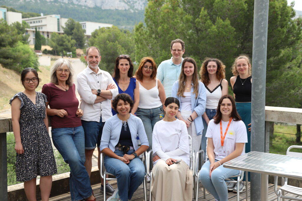
Team & Collaborations
We collaborate closely with partners in immunology, neurobiology, and computation to connect mechanisms at multiple scales—from transcriptional networks to organ morphogenesis.
- Lionel Spinelli, CB2M, CIML, Marseille, France
- Denis Puthier, TAGC, Marseille, France
- Andreas Diefenbach, Charité Berlin, Germany
- Alexandre Pattyn, Montpellier Neuroscience Institute, France
- Léo Guignard, IBDM, Marseille, France
- Rachel Golub, Pasteur Institute, Paris, France
- Christelle Harly, UMR_S 1307 Cancer Research Center and Integrated Immunology Nantes Angers
- Andrea Brendolan, IRCCS Ospedale San Raffaele, Milan, Italy
- Fabio Santori, University of Georgia, USA
- Heather Etchevers, IMM, Marseille, France

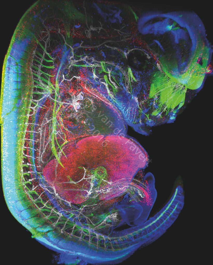
“Immunofluorescent staining of a complete mouse E13.5 embryo reveals neurons (labeled with BetaIII Tubulin), macrophages and lymphatic endothelial cells in red (labeled with Lyve-1), the nuclei of specific neurons and lymphatic endothelial cells are marked in blue (Prox1 labeling) and the vascular network in white (Pecam-1 labeling). The eye of the embryo and the nasal neurons can be seen in green. On the left, the spinal cord is marked in green. The vertebrae are visible through the marking of the dorsal root ganglia in green, and blood vessels marked in white. The heart is visible just above the liver which is labeled with LYVE-1 antibody. The embryonic intestine, not far from the liver, is visualized by the innervation of neurons in green. be modified by changing certain dietary factors affecting cell differentiation. These visualizations are therefore intended to analyze the movements of cells involved in the formation of the lymphatic system.” Image Serge van de Pavert/CIML.
Projects
Project: SensoSkin Apical extracellular matrix (aECM) shapes and protects the epithelium in all organisms. We previously showed that like […]

Amidex Project: ELNINo We analyze how neuronal signals integrate with immune and stromal programs to orchestrate ganglion organogenesis. In […]

Project: CholControl You are what your mother ate Transgenerational impact of maternal diets via the regulation of RORγt activity […]


