Publication: Three-Dimensional Imaging of Macrophages in Complete Organs.
Published in: Methods Mol Biol 2024 ; 2713(): 297-306
Authors: Siret C, van de Pavert SA
Summary
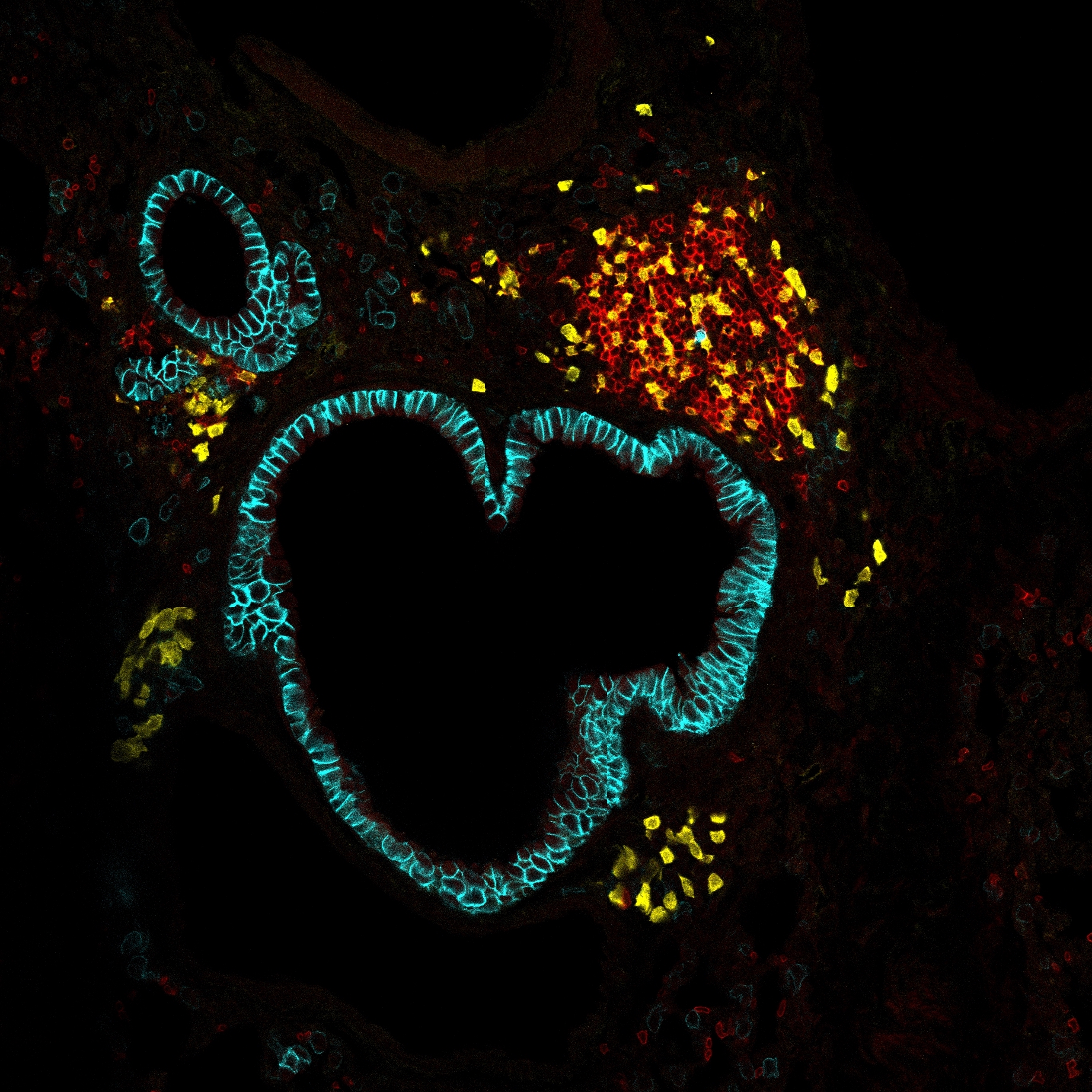
The introduction of the light-sheet microscope has facilitated the analysis of complete tissues for the presence of all cells and their location in relation to their niche. This contributes to a better understanding of cellular locations and interactions in organs. In the last decade, many new and improved protocols have been published which are essential to improve staining and visualization of the immune-fluorescence within different tissues. In this article, we will discuss two main protocols we have used to visualize tissue-resident macrophages.
Link to Pubmed [PMID] – 37639131
Link to HAL – amu-04948583
Link to DOI – 10.1007/978-1-0716-3437-0_20

